






ECG, 2D echo and TMT are often spoken of as though they are interchangeable, or as though doing all three gives a complete answer. They are not interchangeable. Each measures something different, each misses something different, and the single most important fact about the ECG is that a normal result does not rule out coronary artery disease.
This guide explains what each test measures, what it cannot show, and how a doctor decides which one applies.
The Short Version
| Test | What it measures | Takes | Key limitation |
|---|---|---|---|
| ECG | Electrical activity of the heart, at rest | A few minutes | Can be completely normal in significant coronary disease |
| 2D Echo | Structure and pumping function, using ultrasound | 20–30 minutes | Shows the consequences of disease, not the artery blockages themselves |
| TMT (stress ECG) | Electrical changes while the heart works harder | 30–45 minutes | Not suitable for everyone; results can be equivocal |
| Holter | Rhythm over 24–48 hours of normal life | Worn 1–2 days | Only captures what happens while it’s worn |
| CT coronary angiography | The coronary arteries themselves, using CT and contrast | 15–30 minutes | Involves radiation and iodinated contrast |
| Invasive coronary angiography | Arteries directly, via a catheter | About an hour | Invasive; usually when intervention is likely |
ECG (Electrocardiogram)
What it is: Electrodes placed on the chest, arms and legs record the heart’s electrical activity over a few seconds, printed as a tracing.
What it shows well:
- Heart rhythm and rate, including atrial fibrillation and other arrhythmias present at that moment
- Evidence of a heart attack happening now, or one that happened in the past
- Signs of thickened heart muscle, often from long-standing high blood pressure
- Certain electrolyte disturbances and drug effects
What it misses:
- Coronary artery narrowing that is not currently causing a problem. This is the critical point. A person can have significant blockages and a completely normal resting ECG, because at rest the heart is getting enough blood.
- Rhythm problems that come and go, since the recording lasts only seconds
- Valve disease and most structural problems
When it is used: as the first test in almost any cardiac assessment, in emergency chest pain evaluation, before surgery, and as part of routine checkups from middle age.
2D Echocardiogram
What it is: An ultrasound of the heart. A probe on the chest produces moving images of the chambers, valves and the muscle in action.
What it shows well:
- Ejection fraction – the proportion of blood the left ventricle pushes out with each beat, the central measure of pumping function
- Valve structure and function: narrowing (stenosis) and leakage (regurgitation)
- Chamber sizes and wall thickness
- Fluid around the heart
- Regional wall motion abnormalities, where part of the muscle moves poorly, often indicating previous damage
- Congenital structural abnormalities
What it misses:
- The coronary arteries themselves. An echo can show the consequences of poor blood supply, but it does not visualise the blockages.
- Intermittent rhythm problems
When it is used: breathlessness, murmurs, suspected heart failure, after a heart attack, valve follow-up, and before certain treatments.
TMT (Treadmill Test / Stress ECG)
What it is: An ECG recorded continuously while you walk on a treadmill at progressively increasing speed and incline, with blood pressure monitored throughout. The idea is to see what happens when the heart is asked to work harder.
What it shows well:
- Electrical changes suggesting that part of the heart muscle is not receiving enough blood under stress
- Exercise capacity, which is itself a useful prognostic measure
- Blood pressure and rhythm response to exertion
- Whether symptoms are reproduced by exertion
What it misses or cannot do:
- It is not suitable for people who cannot walk adequately on a treadmill
- It can be difficult to interpret when the resting ECG is already abnormal
- Results can be equivocal, requiring a further test
Preparation usually involves: comfortable clothes and walking shoes, a light meal a few hours before rather than a heavy one, and medication guidance from your doctor, since some heart medicines affect the result. Never stop a prescribed medicine on your own before a test.
Imaging the Arteries Directly
When the question is specifically whether the coronary arteries are narrowed, the tests above are indirect. Two tests look at the arteries themselves.
CT coronary angiography uses a CT scanner and injected contrast to show whether the coronary arteries are narrowed or blocked. NICE guidance for suspected stable angina recommends offering CT coronary angiography as an investigation. It involves radiation and iodinated contrast, so suitability depends on kidney function and other factors.
Invasive coronary angiography passes a catheter from an artery in the wrist or groin to the heart and injects contrast directly under X-ray. It is the most definitive assessment and allows treatment such as stenting in the same sitting, which is why it is generally reserved for cases where intervention is likely.
NICE also notes that MR coronary angiography should not be offered to diagnose stable angina.
Blood Tests That Go With Heart Tests
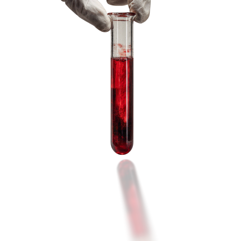
Imaging and electrical tests assess the heart itself. Blood tests assess the risk factors driving disease and anything making symptoms worse:
- Lipid profile: total cholesterol, LDL, HDL, triglycerides
- Fasting glucose and HbA1c: diabetes substantially raises cardiovascular risk
- Complete blood count: NICE specifically notes checking for conditions that can worsen angina, such as anaemia
- Kidney function: relevant to both risk and the safe use of contrast
- Thyroid function: both overactive and underactive thyroid affect the heart
- Troponin: used in the acute setting to detect heart muscle injury, not as a routine screening test
Why “Just Do All of Them” Is the Wrong Approach
Each test answers a specific question, and ordering tests without a question produces three problems:
- False reassurance. A normal ECG and a normal echo can coexist with significant coronary disease. Someone who takes this as an all-clear may ignore genuine symptoms later.
- False alarms. Equivocal findings lead to further scans and procedures that carry their own risks.
- The wrong test for the symptom. Palpitations that come and go need a Holter, not a resting ECG. Breathlessness needs an echo. Exertional chest tightness needs assessment for coronary disease. A bundled package does not make these distinctions.
The sequence is normally: symptoms and history, then examination, then the test that answers the question those raise.
When Chest Pain Is an Emergency
Do not arrange an outpatient test if you have:
- Chest pain or pressure lasting more than a few minutes, or coming and going
- Pain spreading to the arm, jaw, neck or back
- Chest discomfort with breathlessness, sweating, nausea or light-headedness
- Sudden severe breathlessness
- Fainting
Call emergency services or go to the nearest emergency department. Heart attacks are diagnosed with an immediate ECG and blood tests, not with a scheduled appointment. Women, people with diabetes and older adults more often present without classic chest pain, so breathlessness or unusual fatigue should be taken seriously.
Frequently Asked Questions
1. What is the difference between ECG, echo and TMT?
An ECG records the heart’s electrical activity at rest. A 2D echo is an ultrasound showing the heart’s structure and pumping function. A TMT records the ECG while you exercise on a treadmill, to see whether the heart muscle shows signs of inadequate blood supply under stress. They answer different questions and are not interchangeable.
2. Can an ECG be normal even with heart blockage?
Yes, and this is the most important limitation to understand. A resting ECG can be completely normal in someone with significant coronary artery narrowing, because at rest the heart may still receive enough blood. A normal ECG does not rule out coronary artery disease.
3. Which test detects a blocked artery?
Only tests that image the arteries show blockages directly: CT coronary angiography and invasive coronary angiography. ECG, echo and TMT assess the heart indirectly, by looking at electrical activity, structure and function, or the response to stress.
4. Is TMT safe?
It is performed under medical supervision with continuous ECG and blood pressure monitoring, and staff are trained to stop the test if problems appear. It is not suitable for everyone, including people who cannot walk adequately on a treadmill or who have certain acute conditions. Your doctor decides whether it is appropriate for you.
5. What is a normal ejection fraction?
Ejection fraction is the share of blood the left ventricle pumps out with each beat, reported by an echo. Your report will state the measured value against the laboratory’s reference range. Because interpretation depends on your symptoms and history, the number should be reviewed with a doctor rather than compared to a figure found online.
6. Do I need to fast before an ECG or echo?
No. Neither requires fasting. A TMT is usually done after a light meal rather than a heavy one or a long fast. CT coronary angiography and blood tests done alongside may have their own preparation requirements, which the centre will tell you.
7. Which heart test should I do for palpitations?
A resting ECG only captures a few seconds, so it often misses palpitations that come and go. A Holter monitor, worn for 24 to 48 hours during normal activity, is typically used to catch intermittent rhythm problems. Your doctor decides based on how often symptoms occur.
8. Should a healthy person get a heart test in a routine checkup?
An ECG is commonly included in checkups from around age 40 and is reasonable as a baseline. Echo, TMT and CT angiography are not routine screening tests for people without symptoms or risk factors; they are ordered to answer a specific clinical question.
9. What blood tests go with heart tests?
Commonly a lipid profile, fasting glucose and HbA1c, a complete blood count, kidney function and thyroid function. NICE specifically notes checking for conditions that can make angina worse, such as anaemia. Troponin is used in acute settings to detect heart muscle injury, not for routine screening.
10. Can a 2D echo detect a heart attack?
It can show the consequences of one, such as an area of heart muscle that moves poorly, and it helps assess damage afterwards. It is not the test used to diagnose a heart attack as it happens; that relies on an immediate ECG and blood tests such as troponin.
11. How long does each heart test take?
An ECG takes a few minutes. A 2D echo usually takes 20 to 30 minutes. A TMT typically takes 30 to 45 minutes including preparation and recovery. A Holter monitor is worn for 24 to 48 hours. Timings vary by centre.
12. Where can I get heart tests done in Pune?
ECG, 2D echo and TMT all need a centre visit, since they require equipment and supervision. They are available at diagnostic centres across Pune, including Fittr Labs in Kharadi. The blood tests that accompany them, such as a lipid profile and HbA1c, can be collected at home through home sample collection.